Brushings Cytology: Diagnostic Applications in GI, Respiratory, and Urinary Tract Cancers
What is Brushings Cytology?
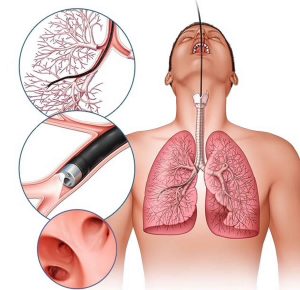
Brushings Cytology is a diagnostic method involving the collection of cells from internal surfaces using a cytology brush during endoscopic procedures. The smears are examined under the microscope for malignancy, infection, and inflammatory changes.
Applies To:
- Colonic Brushings Cytology
- Esophageal Brushings Cytology
- Gastric Brushings Cytology
- Oropharyngeal Brushings Cytology
- Small Bowel Brushings Cytology
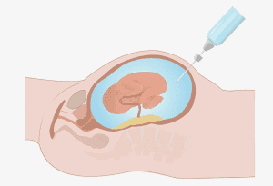
- Urinary Brushings Cytology
Test Usually Includes:
If the brush is submitted in physiological saline, cytocentrifuge preparations can be made. Otherwise, direct smears are prepared from the collected cells.
Patient Preparation:
Obtain informed consent prior to the procedure. No special fasting or dietary restrictions unless specified by the physician.
Specimen Collection:
- Instrument: Flexible fiberoptic bronchoscope or endoscope
- Container: Coplin jar containing 95% ethanol
- Procedure:
- Roll the brush gently over a fully frosted labeled slide.
- Fix the smear immediately in 95% ethanol to preserve cellular details.
- Clearly indicate the brushed site on both the slide and requisition form.
- Send a sterile double-sheathed brush for culture if infection is suspected.
Causes for Sample Rejection:
- Hypocellularity (too few cells)
- Improper fixation or air-drying
- Unlabeled slides
Special Instructions:
Always indicate the exact site brushed and provide full clinical history. Note if special stains for fungi, parasites (e.g., amebas), or viral agents are required.
Uses:
Brushings cytology helps to:
- Diagnose primary or metastatic tumors
- Detect infections from:
- Herpesvirus, Cytomegalovirus (CMV), Measles
- Fungal infections (e.g., Candida, Histoplasma)
- Parasites: Strongyloides, Echinococcus, Giardia, Entamoeba
- Pneumocystis carinii (P. jirovecii)
- Legionella pneumophila (Legionnaires’ disease)
- Perform immunocytochemical staining for tumor or bacterial antigens
Limitations:
Reliable interpretation requires proper fixation. If smears dry out, they become cytologically uninterpretable. However, if air drying is < 30 minutes, rehydration in normal saline for 30 seconds followed by fixation may make the smears usable.
A strong clinical history is crucial. For example, post-tracheostomy atypia may mimic squamous cell carcinoma, requiring clinical correlation.
Additional Notes:
Special cultures and stains may be necessary when infection or inflammation is suspected. Immunostaining enhances the diagnostic accuracy of certain pathologies.
References
- Chambers LA, Clark WE. Acta Cytol, 1986; 30:110–114.
- Cook JJ, Haneman B. Acta Cytol, 1988; 32:461–464.
- Geisinger KR, et al. Cancer, 1992; 69(1):8–16.
- Jeevanandaur V, et al. Gastrointest Endosc, 1987; 33:370–371.
- Melville DM, et al. Am J Clin Pathol, 1988; 41:1180–1186.
- Jacobs, Demott, Finley, et al. Laboratory Test Handbook, Lexi-Comp Inc, 1994.