LE Cell Test (Lupus Test): Procedure, Uses & Limitations
LE Cell Test
Synonyms
LE Prep, LE Preparation, LE Slide Cell Test, Lupus Test
Abstract
The LE Cell Test was once an important diagnostic tool for Systemic Lupus Erythematosus (SLE), but it has now been largely replaced by more sensitive and specific tests like ANA. It remains of historical and academic interest.
Patient Care / Preparation
- Avoid heparin therapy for two days before sample collection.
- High doses of heparin may cause false-negative results.
Specimen
Whole blood
Container
Green top tube (heparin). EDTA tubes may give false negatives.
Collection
- Perform venipuncture.
- Gently invert the tube to mix.
- Transport to the lab within 30 minutes.
Reason to Reject Sample
- Clotted specimen
- Insufficient volume
- Patient currently on high-dose heparin
Reference Range
Negative
Use
Primarily used in the past to help diagnose Systemic Lupus Erythematosus (SLE) and other autoimmune diseases such as lupoid hepatitis.
Limitations
- Less sensitive than fluorescent ANA testing.
- Not specific to SLE—positive results may occur in:
- Drug hypersensitivity
- Rheumatoid arthritis
- Chronic active hepatitis
- Other collagen vascular diseases
- A negative result does not rule out lupus.
- Use of EDTA as anticoagulant may yield false negatives.
Contraindications
- High-dose heparin therapy
- Severe leukopenia or neutropenia
Methodology
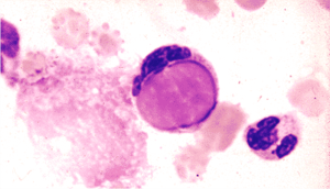
Blood cells are lysed (commonly using glass beads), releasing nuclear material. This interacts with autoantibodies, forming phagocytosed LE cells. Slides are prepared from the buffy coat, stained, and observed for lavender-staining phagocytosed nuclei under a microscope. The presence of extracellular LE material or rosettes is not diagnostically significant.
Additional Information
- SLE primarily affects women and presents with symptoms such as:
- Rash
- Fever
- Arthralgia
- Anemia, leukopenia, thrombocytopenia
- Hypocomplementemia
- LE slide test is positive in only 60–80% of active SLE cases.
- Negative results must be followed up with an ANA test (95% sensitivity).
- Most commonly found antibodies are of the IgG class.
- Drug-induced lupus may also cause positive LE tests; e.g., Diphenylhydantoin (Dilantin®) users.
References
- Dacie JV, Lewis SM. “Demonstration of LE Cells.” Practical Haematology, 7th ed. Churchill Livingstone, 1991.
- Hargraves MM. “Discovery of the LE Cell.” Mayo Clin Proc. 1969.
- Hargraves MM, Richmond H, Morton R. “LE & Tart Cell Presentation.” Proc Mayo Clin, 1948.
- Nakamura RM, Peebles CL, Rubin RL, et al. “Autoantibodies to Nuclear Antigens (ANA).” ASCP Press, 1985.
- Steinberg AD, Gourley MF, Klinman DM. “Systemic Lupus Erythematosus.” Ann Intern Med, 1991.
- Jacobs et al. “Laboratory Test Handbook.” Lexi-Comp Inc, 1994.