Osmotic Fragility Test
Synonyms
Incubated Osmotic Fragility, RBC Fragility, Red Cell Fragility
Specimen
Whole blood
Container
Lavender top (EDTA) or Green top (Heparin) tube
Note: Avoid oxalate or citrate anticoagulants.
Storage Instructions
Refrigerate at 4°C if testing is delayed.
Reason to Reject Sample
- Use of oxalate or citrate anticoagulants
- Hemolyzed or clotted sample
- Sample older than 6 hours
Special Instructions
Test may require advance scheduling.
Reference Range
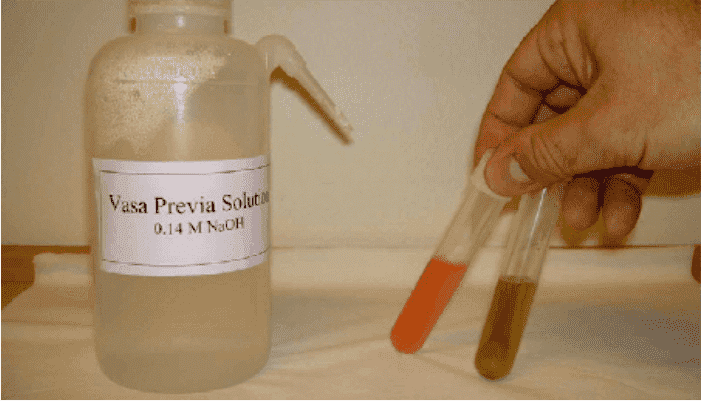
- Hemolysis begins: 0.45% NaCl
- Complete hemolysis: 0.35% NaCl
Use
- Primary use: Suspected hereditary spherocytosis
- Supportive diagnosis for hemolytic anemias (immune or structural)
Limitations
- Iron deficiency anemia, thalassemia, or hemoglobinopathies may alter results
- No test available exclusively for hereditary spherocytosis
- Patient discomfort during venipuncture can impact results
Methodology
Erythrocytes are incubated in varying concentrations of sodium chloride (NaCl). Hemolysis occurs in hypotonic solutions, and the degree of fragility is measured using optical density. Additives like Glycerol and Bis-Tris may increase test sensitivity for hereditary spherocytosis.
Additional Information
Hereditary spherocytosis is often inherited in an autosomal dominant pattern and is linked to mutations in red cell membrane proteins such as spectrin, ankyrin, and protein 4.2. These structural abnormalities result in spherocytes—red cells that are more fragile in hypotonic solutions.
Conditions with Increased Osmotic Fragility
- Hereditary spherocytosis
- Autoimmune hemolytic anemia
- Malaria (in both infected and uninfected cells)
- Multiple sclerosis (as reported in some cases)
Conditions with Decreased Osmotic Fragility
- Iron deficiency anemia
- Thalassemia
- Hemoglobin C disease
- Presence of target cells or hypochromic cells
- Some cases of stomatocytosis
Clinical Note
In hereditary spherocytosis, splenectomy is often therapeutic and can relieve hemolysis. Patients may present with reticulocytosis, elevated indirect bilirubin, gallstones, or even aplastic crises. Molecular diagnostics reveal that most cases involve mutations in spectrin or associated anchoring proteins.
References
- Dacie JV & Lewis SM. Practical Haematology. 7th ed. Churchill Livingstone, 1991:196–200.
- DiPaolo BR, Speicher KD, Speicher DW. Blood, 1993; 82(1):284–91.
- Henry JB et al. Clinical Diagnosis and Management by Laboratory Methods. WB Saunders Co, 1991.
- Palek J. Semin Hematol, 1993; 30(1):1–3.
- Peters LL & Lux SE. Semin Hematol, 1993; 30:85–118.
- Winkelmann JC & Forget BG. Blood, 1993; 81(12):3173–85.
- Jacobs, Demott, Finley, Horvat, Kasten.JR, & Tilzer. Laboratory Test Handbook. Lexi-Comp Inc, 1994.