KOH Preparation: A Simple Diagnostic Test for Fungal Infections

Synonyms
Potassium Hydroxide Preparation
Test Commonly Includes
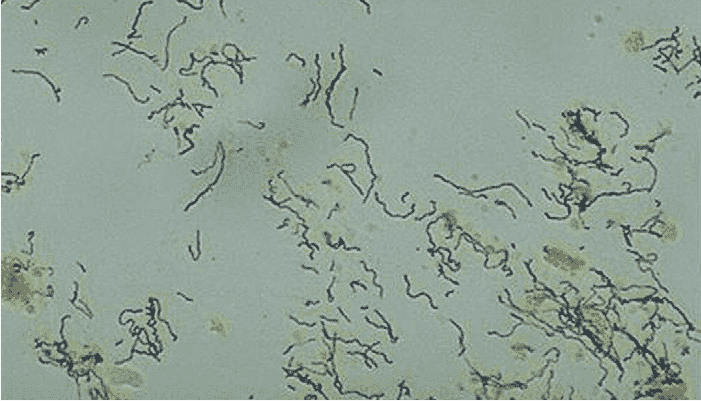
KOH (Potassium Hydroxide) dissolves keratin and other proteinaceous material, allowing fungal elements (hyphae, spores, or yeast) to be seen clearly under a microscope at 10x and 40x magnification.
Patient Care / Preparation
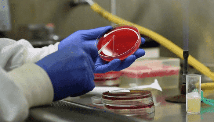
Same as the preparation required for a fungal culture from the same site.
Specimen
Identical to the specimen used for fungal culture. Can include skin scrapings, nail clippings, hair, corneal scrapings, or mucosal secretions.
Container
The same type of container used for culture from the respective collection site.
Special Instructions
Notify the laboratory of the exact specimen source and clinical diagnosis to aid interpretation.
Reference Range
No fungal elements observed.
Use
- To detect fungal infections in skin, nails, and hair (e.g., tinea, onychomycosis, keratomycosis).
- To assist in the diagnosis of fungal infections before initiating antifungal therapy.
Limitations
- KOH preparation is less sensitive than fungal culture.
- A negative result does not rule out fungal infection.
- May require overnight incubation for hard specimens like nails or hair to dissolve keratin adequately.
Methodology
The test typically uses:
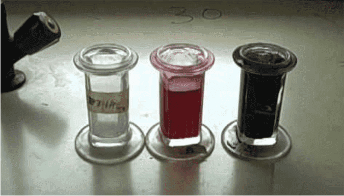
- 10% or 20% Potassium Hydroxide (KOH)
- KOH with 40% Dimethyl Sulfoxide (DMSO) for rapid clearing
- Gentle heating may help in faster hydrolysis of tough debris
Procedure
- Place the specimen on a glass slide.
- Add a drop of 10–20% KOH (optionally with DMSO).
- Gently heat the slide if needed (avoid boiling).
- Place a coverslip and wait a few minutes to clear the background.
- Examine under 10x and 40x microscope objectives.
Microscopic Findings
- Dermatophytes: Long, branching hyphae
- Yeasts (e.g., Candida): Budding cells with or without pseudohyphae
- Acanthamoeba cysts: Sometimes visible (fluorescent stains like Calcofluor White preferred)
Additional Notes
- KOH smears are especially useful in diagnosing keratomycosis and tinea infections.
- In corneal infections, KOH smears may be more sensitive than culture due to presence of dead fungal elements.
- Hair fragments or “black dots” under dermoscopy or Wood’s lamp should be examined under KOH.
- Topical steroids should be avoided before confirming the diagnosis to prevent masking symptoms.
References
- Stein DH, “Superficial Fungal Infections,” Pediatr Clin North Am, 1983
- Ishibashi Y et al., “Direct vs Culture for Keratomycosis,” Am J Ophthalmol, 1987
- Gray LD & Roberts GD, “Systemic Fungal Disease Diagnosis,” Infect Dis Clin North Am, 1988
- Krowchuk DP et al., “Tinea Capitis Identification,” Pediatrics, 1983
- Jacobs, Demott, Finley et al., “Lab Test Handbook”, Lexi-Comp Inc, 1994