Darkfield Microscopy for Syphilis: Diagnosis Using Treponema pallidum Examination
Synonyms
Syphilis Darkfield Microscopy, Treponema pallidum Darkfield Examination
Test Commonly Includes
Cleansing of chancre or condyloma, collection of exudate or serum, and immediate examination under a darkfield microscope.
Abstract
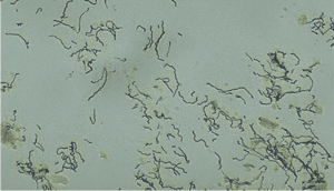
Treponema pallidum is a thin spirochete not visible under standard light microscopy. Darkfield microscopy is essential for detecting this organism in moist lesions such as chancres (primary syphilis) and condylomata lata (secondary syphilis).
Patient Care & Preparation
- Clean lesion with a saline-moistened swab to remove exudate and contaminants
- Collect subsurface serum using a small pipette or by pressing a slide directly against the lesion
- Examine immediately; mobility of organisms diminishes rapidly with temperature drop
Specimen
Moist serum from the base of an unhealed, untreated chancre or condyloma. The freshest and youngest lesion yields the best diagnostic results.
Specimen Rejection Criteria
- Dry or bloody specimens
- Ointment contamination
- Previously treated or healing lesions
Reference Range
Treponema pallidum appears as a tightly coiled, motile organism, 0.10–0.18 µm wide and 6–20 µm long (about 1–1.5× the diameter of an RBC).
Use
To confirm the presence of spirochetes in lesions suspected of being syphilitic. Especially valuable in primary syphilis diagnosis.
Limitations
- Darkfield exam is unreliable for oral or rectal lesions due to presence of nonpathogenic spirochetes
- Ineffective for dry, bloody, or old lesions
- Specimens must be examined within 15 minutes
Contraindications
- Do not perform after antibiotic treatment
- Organisms are rapidly killed and become undetectable post-treatment
Methodology
Darkfield microscopy visualizes live, motile spirochetes. T. pallidum shows rapid, corkscrew-like movement and distinctive bending or flexing at 90° angles.
Additional Information
- More common in primary syphilis lesions than in secondary
- Cannot be cultured in vitro
- Fluorescent antibody techniques are an alternative when darkfield is not available
References
- Hook EW 3d & Marra CM, N Engl J Med, 1992
- Fitzgerald TJ, Manual of Clinical Microbiology, 5th ed., ASM, 1991
- Larsen SA, Clin Lab Med, 1989
- Jacobs, Demott, Finley, et al., Laboratory Test Handbook, Lexi-Comp Inc, 1994